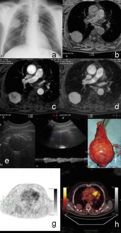
Figure 3.
SFTP in a 63-year old man. This was an incidental finding of a chest radiograph obtained in a patient with cardiac arrhythmia. PA chest radiograph (a) demonstrating a rounded mass in the right middle hemi-thorax. The lower edge of the lesion is well-defined, but its right-superior border is ill-defined suggesting a pleural origin. Corresponding axial T1-weighted MRI shows a mass of intermediate to high signal intensity (b), while multiphase gadolinium-enhanced T1-weighted image demonstrates early heterogeneous enhancement (c). During the 2-min-late phase, the lesion showed a more homogeneous contrast enhancement than in the arterial phase (d). Corresponding sonographic scan (e) showing a hypoechoic rounded mass with regular margins. Peripheral signals at colordoppler show the presence of a capsular vascularization corresponding to the real anatomy of the lesion (f ). The doppler signals inside the mass are due to centripetal blood vessels moving from the vascular hilum. PET (g) and PET-CT fusion (h) images of the same patient demonstrate a low activity mass.

