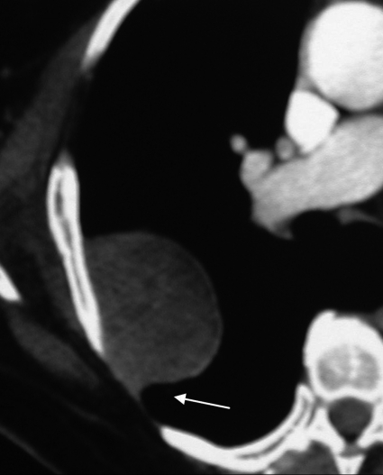
Figure 9.
Benign SFTP in an asymptomatic 71-year old man. Chest enhanced CT scan targeted to the visualization of the lesion (mediastinal window) demonstrates a homogeneous oval lobular soft tissue mass abutting the parietal pleura. Although the lesion forms an acute angle with the pleura layers, a smooth tapering margin (arrow) is also seen.

