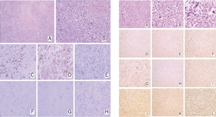
Figure 3.
Immunohistochemical staining of the resected lesion. Left column (right lung lesion): H&E stain of tumor cells; 100× (A) and 200× (B) magnification. Tumor cells were positive for cytokeratin (C) and vimentin (D). Epithelial membrane antigen (E) was focally positive while cytokeratin 7 (F), CD20 (G), and desmin (H) were negative. Right column (intestinal lesion): H&E stain of tumor cells; 100× (A), 200× (B), and 400× (C) magnification. Tumor cells were negative for CD117 (D), CD×2 (E), and CD20 (H). Cytokeratin (F), epithelia/membrane antigen (I), S-100 (J), TTF-1 (K), and vimentin (L) were positive. Cytokeratin 7 (G) was focally positive.

