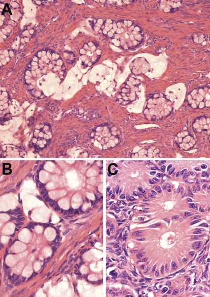
Figure 1.
(A)Primary tumor (left ovary): small round glands sometimes floating in lakes of mucin typified the primary tumor (hematoxylin and eosin, 100×). (B) Glands with small minimally atypical basal nuclei predominated in this tumor (hematoxylin and eosin, 400×). (C) In places there was moderate cytological atypia and some fusing of glands (hematoxylin and eosin, 400×).

