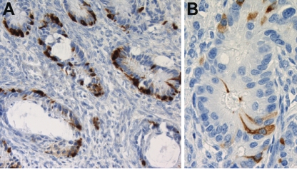
Figure 3.
Immunohistochemistry in primary tumor and metastatic tumor. (A) Metastatic tumor (right ovary): cells staining positively for chromogranin were present in most glands (200×). (B) Primary tumor (left ovary): scattered cells stained positively with synaptophysin, in a gland with cytological atypia (400×).

