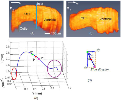
Fig. 4.

Boundary segmentation of chicken embryonic OFT walls: (a) Structural image; (b) boundary segmentation; (c) segmented boundary curve; (d) region within the curve (geometrical center of the region serves as blood vessel center point).

Boundary segmentation of chicken embryonic OFT walls: (a) Structural image; (b) boundary segmentation; (c) segmented boundary curve; (d) region within the curve (geometrical center of the region serves as blood vessel center point).