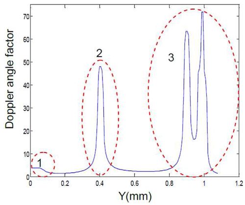
Fig. 5.

Doppler angle factor (DF) distribution at different positions along the chicken embryo OFT. Region 1: OFT near the arterial system; region 2: bended region of OFT; region 3: ventricle. Doppler angle factors in region 1 and 3 are inaccurate because the boundaries of the OFT in the structural image are not clear (Section 3.3). Doppler angle factors in region 2 are not useful because the Doppler angles here are close to π/2 (Section 3.4).
