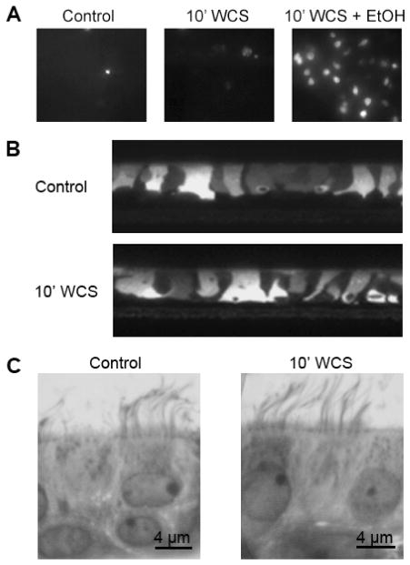
Figure 2.
WCS does not affect HBEC integrity. Viability (A). All cultures (control cultures, cultures 10 min after undiluted WCS exposure and WCS-exposed cultures after addition of 70% ethanol) were stained with 5 μM propidium iodide, which fluoresces upon binding to DNA of dead cells (all images are representative from n = 4 cultures per group). Integrity (B). Calcein-stained live HBECs imaged by confocal microscopy pre- (top) and post- (bottom) 10-min WCS exposure (all images are representative from n = 4 cultures per group). Morphology (C). Control HBECs and HBECs exposed to WCS for 10 min were fixed in 1% osmium tetraoxide suspended in perfluorocarbon and stained with Richardson’s stain before being imaged by light microscopy with 100× lens.

