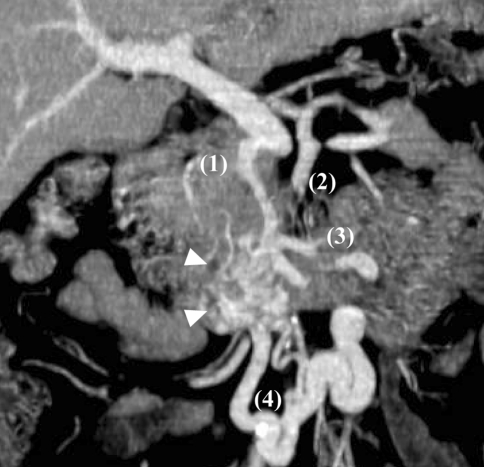
Fig. 1.
CT scan image by multiplanar reconstruction CT. The image revealed the duodenal varices (arrowhead) with three afferent vessels: posterior superior pancreaticoduodenal vein (1), dorsal pancreatic vein (2), and superior mesenteric vein via jejunal vein (3). Two efferent vessels draining through the veins of Retzius (4) were also depicted

