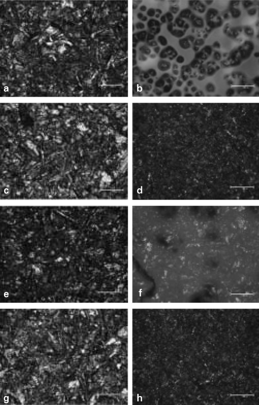
Fig. 4.
Optical microscope photomicrographs of surface treated metal specimens (× 200). a. CS, b. CM, c. CT, d. CC, e. TS, f. TM, g. TT, h. TC. CS, TS, CT and TT group showed irregular aspect with many undercuts. The milky thick membrane was found in CM and TM group. CC and TC group showed less irregular aspect with more undercuts than CS and TS groups and also had milky thin membrane.

