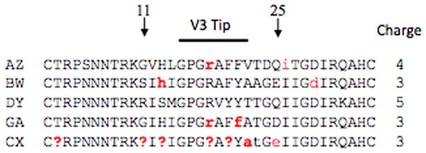
Figure 1. V3 loop consensus sequences.

The 35 consensus amino acids that span the V3 loop domain along with the overall charge of the domain are shown for each patient. Conserved positions, those that occur at a level of 95% or more, are shown in black and variable positions are shown in red. Lower case letters indicate a position that is conserved at a level of 51–94%, whereas a question mark indicates a position where no majority is observed.
