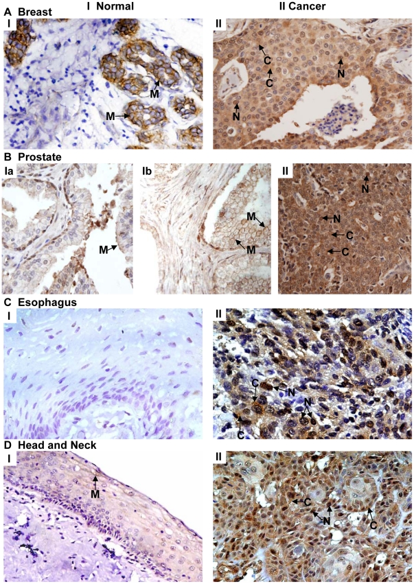
Figure 1. Immunohistochemical analysis of Ep-ICD expression in epithelial cancers and normal tissues.
The representative photomicrographs depict Ep-ICD immunostaining in normal and cancer tissues. Panel A shows predominant membrane localization of Ep-ICD and no nuclear staining in the normal breast tissue (I), while the cancer tissue shows nuclear and cytoplasmic Ep-ICD accumulation (II). Panel B shows low level of membrane Ep-ICD in the epithelial cells and the basal cells show some nuclear staining in the normal prostate tissue (Ia) and in benign prostate hyperplasia (B, Ib), while the cancer tissue shows intense cytoplasmic and nuclear staining (II). Panel C shows no detectable Ep-ICD staining in the normal esophageal tissue (I), while the ESCC shows intense nuclear and cytoplasmic immunostaining (II). Panel D depicts head and neck normal mucosa showing faint membrane Ep-ICD (I), while the HNSCC shows intense nuclear and cytoplasmic immunostaining (II). Original magnification ×400.

