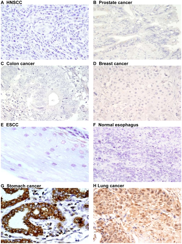
Figure 4. Ep-ICD immunohistochemical analysis in epithelial cancers control tissues.
The negative and positive control photomicrographs are shown. Ep-ICD negative controls for HNSCC (A), prostate cancer (B), colon cancer (C), breast cancer (D), ESCC (E), and normal esophagus (F); panels G and H are positive controls for Ep-ICD staining. Original magnification ×400.

