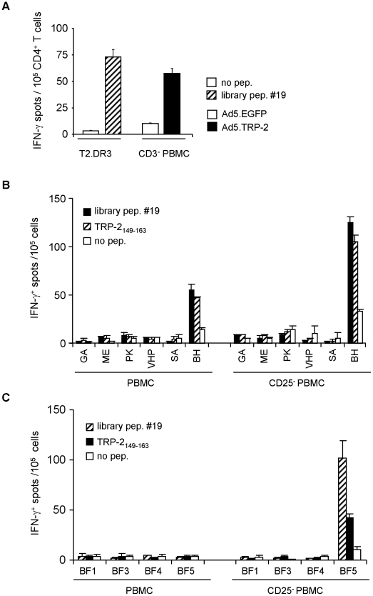
Figure 3. CD4+ T cells from melanoma patients respond to the HLA-DRB1*0301-restricted epitope TRP-2149–163.
A, PBMC from a HLA-DRB1*0301+ healthy donor were primed in vitro with autologous DC pulsed with library peptide #19. After two rounds of in vitro restimulation, CD4+ T cells were tested against T2.DR3 target cells pulsed with library peptide #19 and for recognition of autologous CD3+-depleted PBMC (CD3− PBMC) infected with Ad5.TRP-2 or with control virus. One representative experiment out of two is presented. B, Total PBMC (left panel) or CD25+-depleted PBMC (right panel) from six HLA-DRB1*03+ melanoma patients and C, four HLA-DRB1*0301+ healthy donors were incubated in vitro with library peptide #19. After 17 days, cells were tested by IFN-γ ELISpot assay for their reactivity against library peptide #19 or the epitope TRP-2149–163. All determinations were performed at least in duplicates. Data are presented as mean numbers of IFN-γ spots per 105 cells. Error bars show standard error of the mean.

