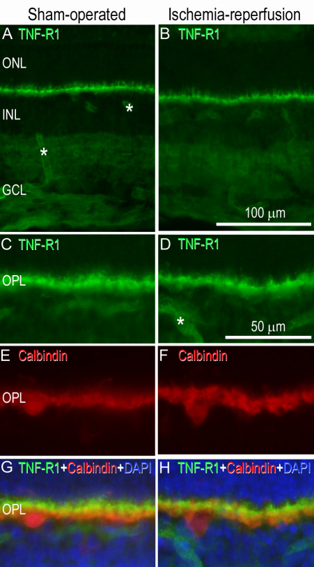
Figure 8.
Tumor necrosis factor (TNF)-R1 immunofluorescence staining in the neuroretina. Representative examples of (A) a sham-operated eye and (B) the fellow eye subjected to ischemia and 12 h of reperfusion showing TNF-R1 staining in the outer plexiform layer of the neuroretina. C-H: The smaller pictures are enlargements showing double staining for (C-D) TNF-R1 and (E-F) calbindin antibodies. Co-localization (G-H) could be seen in the horizontal cells. G-H: DAPI showed staining of the nucleus. Asterisks indicate labeled blood vessels. The original and enlarged images are from separate sections. Abbreviations used in the figure are outer nuclear layer (ONL), inner nuclear layer (INL), ganglion cell layer (GCL) and outer plexiform layer (OPL).

