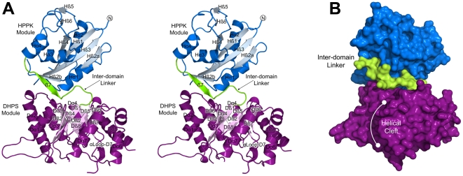
Figure 4. The overall structure of the HPPK-DHPS bifunctional enzyme from Francisella tularensis.
(A) A stereo view of the overall fold and domain organization showing the secondary structure elements within each module. Each element is labeled with the prefixes ‘H’ and ‘D’ to reflect their locations in the HPPK (blue) and DHPS (purple) domains, respectively. The N- and C-termini and the linker region (green) are labeled. Note that helix Dα8 in the canonical DHPS TIM-barrel is missing. (B) A surface representation of the view shown in (A) that highlights the position of the domain linker and the cleft within the DHPS module corresponding to the missing Dα8 TIM-barrel α-helix.

