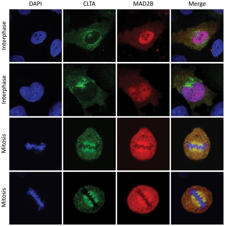
Figure 2. MAD2B and CLTA co-localize at the mitotic spindle.
Cells were transiently transfected with CLTA-GFP (green) and MAD2B-mRFP (red) constructs. DAPI staining (blue) marks the position of the nuclei and chromosomes. Yellow staining in the overlay indicates co-localization of MAD2B and CLTA. The upper two panels show cells in interphase, the lower two panels show cells in mitosis.

