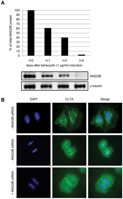
Figure 3. Cellular re-distribution of CLTA after MAD2B knockdown.
(A) HEK293/T-REx cells were stably transfected with a pSUPERIOR-MAD2B siRNA construct (see Materials and methods). Subsequently, tetracyclin was added and cells were collected after 0, 1, 2 and 4 days, lysed and subjected to western blot analysis using anti-MAD2B and anti-γ-tubulin (control) antibodies. (B) HEK293/T-REx/pSUPERIOR-MAD2B cells were grown with or without tetracyclin (+/− MAD2B siRNA, respectively). Endogenous CLTA proteins were detected using an anti-CLTA antibody (green). DAPI staining (blue) was used to mark nuclei and chromosomes. In the presence of MAD2B CLTA localizes to mitotic spindles during mitosis, whereas after MAD2B depletion CLTA is re-distributed throughout the cell.

