Figure 2.
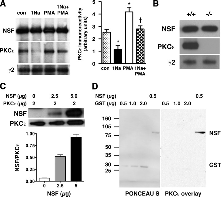
PKCε interacts directly with NSF in a complex containing GABAA receptor γ2 subunits. A, The left panel is a representative experiment showing proteins immunoprecipitated from HEKGR-AS-PKCε cell lysates with anti-NSF antibody and detected by Western blot analysis with anti-NSF, anti-PKCε, or anti-γ2-subunit antibodies. Data are quantified in the right panel, showing that, after 45 min of treatment, 1 μm 1Na-PP1 decreased and 1 μm PMA increased the amount of PKCε coimmunoprecipitated with NSF [n = 3; *p < 0.05 compared with control (con) and †p < 0.05 compared with PMA by Newman–Keuls post hoc test]. B, Representative Western blots with anti-NSF, anti-PKCε, and anti-γ2-subunit antibodies of brain lysates from Prkce+/+ and Prkce−/− mice after immunoprecipitation with anti-NSF antibody. C, The top panel shows representative Western blots from a pull-down assay using recombinant NSF and purified FLAG-PKCε (2 μg) immobilized on anti-FLAG antibody-conjugated agarose. The amount of NSF pulled down with immobilized PKCε increased linearly with the amount of NSF added to the assay (bottom panel; n = 4). D, Representative overlay assay showing Ponceau S-stained-NSF and GST immobilized on a nitrocellulose membrane after SDS-PAGE (PONCEAU S; left) and PKCε immunoreactivity detected using an anti-PKCε antibody (PKCε overlay; right) after incubation of the membrane with recombinant PKCε. This experiment were repeated three times with similar results. Error bars indicate SEM.

