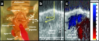
Figure 2.
Intraluminal Doppler OCT imaging using exogenous scattering contrast agents. Panel (a) is a still frame taken from Video 1 and shows the microinjection technique. Panel (b) is a structural OCT image of the heart during systole a few minutes after injection of contrast agent into the thorax. Panel (c) has the Doppler OCT image superimposed on the structural OCT image. The Doppler angle is estimated to be 110 deg. Also clearly seen in panel (c) is the Doppler signal generated by the ventral wall.

