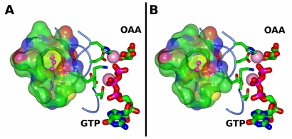
Figure 3. Structural models of cytosolic PEPCK.
Based on previously published structural models of (A) rat cytosolic PEPCK (isoleucine 267) (PDBID 3DT7 [33]) and (B) human cytosolic PEPCK variant rs8192708G (valine 267) (PDBID 1KHB [31]), the hydrophobic pocket to which the residue at position 267 belongs is rendered as a semi-transparent molecular surface colored by atom type (carbon, green; oxygen, red; nitrogen, blue; sulfur, yellow). The van der Waals surface for isoleucine (A) and valine (B) is rendered as a dotted yellow surface. The metal ions are rendered as pink spheres, the catalytic and metal binding residues are rendered as thin cylinders colored by atom type and the substrates oxaloacetate (OAA) and guanosine triphosphate (GTP) are rendered as thick sticks, colored by atom type and labeled.

