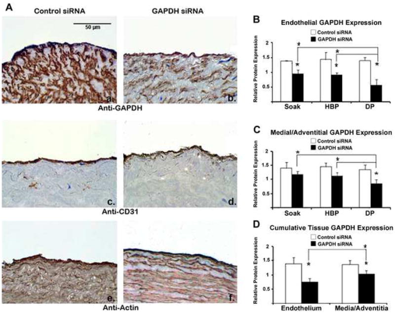
Figure 3.

Endothelial GAPDH protein knockdown exceeds medial and adventitial GAPDH knockdown and is increased using distending pressure transfection. (A) Immunohistochemistry demonstrates GAPDH knockdown within all tissue layers after distending pressure (DP) transfection with GAPDH siRNA (panel b) as compared to vein segments treated with control siRNA (panel a). Protein knockdown is specific to GAPDH as CD31 (panels c,d) and actin (panels e,f) levels are preserved. Quantitation demonstrates (B) greater GAPDH knockdown in the endothelium using distending pressure as compared to hyperbaric (HBP) and non-pressurized (soak) transfection. (C) Medial/adventitial GAPDH levels were significantly reduced after distending pressure transfection, but not with hyperbaric or non-pressurized transfection. (D) Cumulative analysis of all transfections revealed greater knockdown in the endothelium as compared to the media/adventitia. n = 3 vein segments per condition. Micrographs (×400) correspond to one representative image of three experiments performed. (* denotes P < 0.05 for comparisons).
