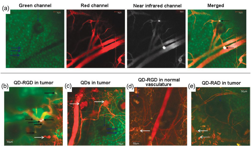
Figure 3.
Direct visualization of QD800-RGD binding to tumor vessel endothelium and controls in a living mouse ear tumor model using intravital microscopy. (a) Each panel displays different output channels of the identical imaging plane along the row. In the green channel, individual enhanced green fluorescent protein (EGFP)-expressing cancer cells are visible, while the red channel shows the tumor’s vasculature via injection of Angiosense dye. The NIR channel shows intravascularly administered QDs that remain in the vessels. Binding events are visible by a bright white signal. These are demarcated by arrows in the rightmost merged image in which all three channels have been overlaid. (b) Merged image of a different mouse using QD800-RGD. Individual cells are not generally visible. (c–e) Typical images without binding in each control condition: (c) Tumor neovasculature containing unconjugated QDs, (d) normal vasculature containing QD800-RGD, and (e) tumor neovasculature containing QD800-RAD. Reprinted with permission from Ref. [76]. Copyright 2008, American Chemical Society.

