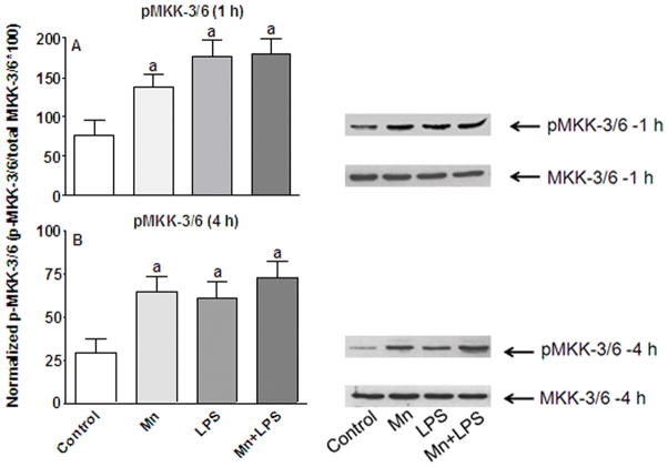
Figure 1.
Effects of Mn and/or LPS on MKK-3/6 activity. Shown are quantification and representative western blots of phosphorylated MKK-3/6 (pMKK-3/6; Ser 189) and total MKK-3/6 in N9 microglia following exposure to vehicle, 250 μM Mn, 100 ng/ml LPS, or 250 μM Mn + 100 ng/ml LPS for 1 (A) and 4 (B) h. Densitometric data were normalized as a ratio of phosphorylated to total MKK-3/6 protein. All data points represent means ± SEM from at least 3 independent experiments. Data were analyzed with ANOVA and means were separated using Fisher’s LSD multiple comparison post hoc test. a Letters denote statistically significant difference from control at p ≤ 0.05.

