
Fig 5.
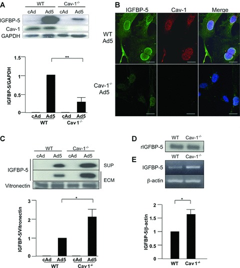
IGFBP-5 levels in lysates, supernatants and ECM of WT and Cav-1−/− lung fibroblasts. Cultured WT and Cav-1−/− fibroblasts were infected with cAd and Ad5 at an MOI of 50. Lysates, supernatants and ECM were harvested after 48 hrs (A) or 72 hrs (C). Levels of IGFBP-5 were analysed by immunoblotting of lysates (A), supernatants (SUP) and ECM (C). GAPDH and vitronectin were used as loading controls for lysates and ECM, respectively. For the supernatant, an equivalent amount of total protein was loaded in each lane. Graphical summary of IGFBP-5 expression in lysates (A) and ECM (C) from WT and Cav-1−/− fibroblasts. The unpaired t-test was used for statistical analysis. Horizontal bars indicate mean values of three independent experiments for (A) and two independent experiments for (C). *P < 0.05, **P < 0.01. (B) IGFBP-5 and Cav-1 localization was examined by immunocytostaining. WT and Cav-1−/− fibroblasts were cultured on cover slips coated with type I collagen. After infection with Ad5 for 48 hrs, cells were stained for IGFBP-5 (green) and Cav-1 (red). Hoechst (blue) was used to identify nuclei. Magnification of images was 600× on a confocal microscope. Scale bars = 20 μm. (D) Western blot analysis of rIGFBP5 levels following 1 hr incubation with media conditioned by WT and Cav-1−/− fibroblasts. (E) Steady-state mRNA levels of mouse IGFBP-5 in NT, WT, and Cav-1−/− fibroblasts. β-actin was used as a control. Normalized IGFBP-5 mRNA levels in WT were arbitrarily set at 1. Horizontal bars indicate mean values of three independent experiments. The unpaired t-test was used for statistical analysis.