Figure 3.
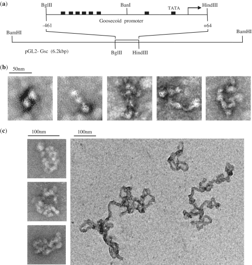
Electron microscopy of PRH with short and long DNA fragments. (a) A schematic of the DNA fragments used in the EM experiments shown here. The top line shows a 525-bp DNA fragment carrying Goosecoid sequences from –461 to +64 relative to the transcription start point obtained by digestion of pGL-Gsc with BglII and HindIII. The bottom line shows the same plasmid linearized by digestion with BamHI to produce a 6.2 kbp DNA fragment with centrally located Goosecoid promoter sequences. (b) The 525-bp DNA fragment shown in (a) was incubated with PRH (1 µM) in binding buffer for 20 min at 4°C. The complexes formed were deposited on a carbon-coated copper EM grid and stained with 1% uranyl acetate for transmission EM. (c) The experiment in (b) was repeated with the 6.2 kbp DNA fragment shown in (a).

