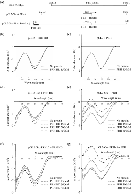
Figure 4.
Linear dichroism indicates DNA compaction by PRH in solution. (a) A schematic of the DNA fragments used in LD experiments. The top line shows pGL2-basic linearized by digestion with BamHI. The middle line shows pGL2-Gsc linearized using the same restriction enzyme. The open rectangle and arrow represent the Goosecoid promoter sequences and Goosecoid transcription start point, respectively. The bottom line shows the pGL2-Gsc-PRHx5 plasmid linearized with SalI. The filled rectangle represents the five PRH binding sites cloned into the BamHI site of pGL2-Gsc to produce this construct. (b–g) LD of the plasmid DNAs shown above (5 nM) with PRH-HD (b, d and f) or the full-length PRH protein (c, e and g).

