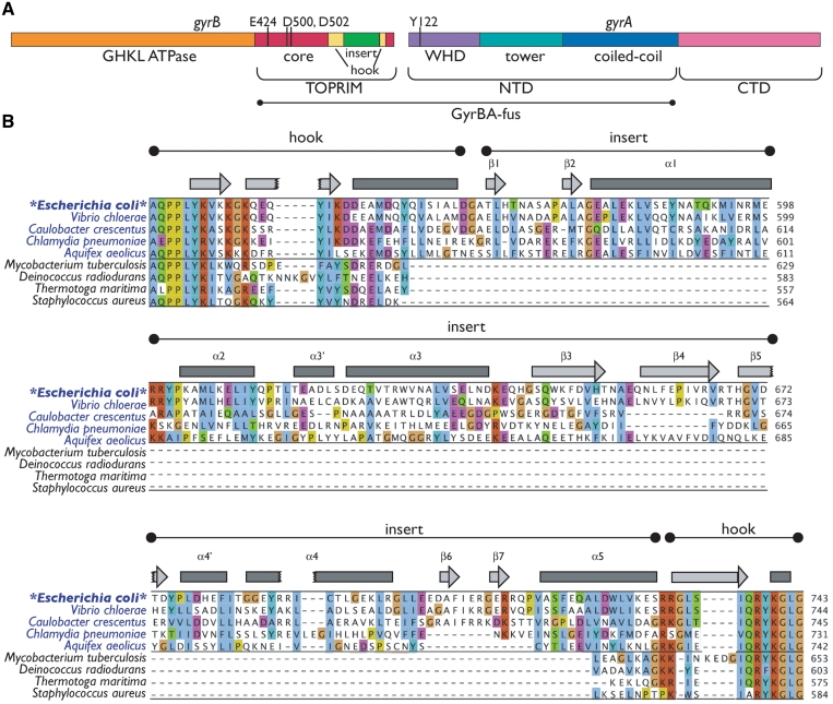
Figure 1.
Escherichia coli gyrase primary and secondary structure. (A) GyrA and GyrB domain organization. Catalytic modules of E. coli gyrase are indicated as bars. The regions encompassed by the GyrBA fusion are indicated. (B) Alignment of the GyrB insert region. Selected GyrB genes were aligned using MUSCLE (68,69). Organisms with GyrB genes containing an insert are shown in blue; those with GyrB genes lacking inserts, in black. Regions corresponding to the hook and insert are indicated, as are secondary structure elements as identified in the structure (bars—α-helices; arrows—β-sheets). Insert elements are numbered by secondary structure element as in Figure 3B. Amino acid numbering is at right. The alignment is colored by ClustalX score within the two major subgroups (GyrBs with and without inserts). Note the poor conservation of the insert compared to the hook region. This figure was prepared using Jalview (70).

