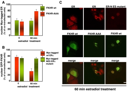
Fig. 3.
Estradiol simultaneously induces nuclear export of ER and FKHR in MCF-7 cells. Quiescent MCF-7 were co-transfected along with the indicated constructs and then left unstimulated or stimulated for 60 min with 10 nM estradiol. Graphs in A represent the nuclear score of Myc-tagged ER in MCF-7 cells co-expressing either GFP-FKHR wt (green bars) or the GFP-FKHR-AAA mutant (red bars). Graphs in B represent the nuclear score of GFP-FKHR in MCF-7 cells co-expressing either Myc-tagged ER alpha wt (red bars) or the Myc-tagged ER alpha NES mutant (green bars). Images of confocal microscopy analysis were captured and are shown in C. They represent the staining of Myc-tagged ER alpha in MCF-7 cells expressing GFP-FKHR wt (left in green), or the GFP-FKHR-AAA mutant (middle in green) and treated for 60 min with 10 nM estradiol. Right panels represent the staining of Myc-tagged ER alpha NES mutant (red) in MCF-7 cells co-expressing GFP-FKHR wt (green) and treated for 60 min with 10 nM estradiol. Merged images are shown at the bottom. Bar, 5 μm. For experimental details see also Lombardi et al. 2008 in refs

