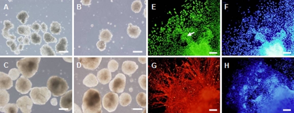
Fig. 9.
Neual differentiation of HN4 in vitro. a–d Floating EBs of HN4 hES cells at 6, 24 h, 4 and 14 days. e–f Differentiated cells at day 2 after plating. e Nestin positive cells. Nestin stains neural tube—like structures (pointed by white arrow). g–h Differentiated cells at day 18 after plating. g β-III-tubulin positive cells. f and h Nuclei were stained with DAPI (blue). Scale bar = 200 μm

