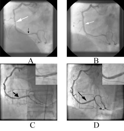
Fig. (1).
A. Coronary angiogram five years before showing the lesion in the middle RCA (white arrow). The distal RCA is normal, B. The artery after stenting of the lesion (white arrow), C. Spiral dissection in the distal RCA (black arrow) manifested with inferior acute myocardial infarction. The dissection is also depicted in magnification on the upper right corner, D. Distal RCA after stenting of the dissection (black arrow). The restoration of vessel patency after successful stenting is also depicted in magnification on the upper right corner.

