Figure 6.
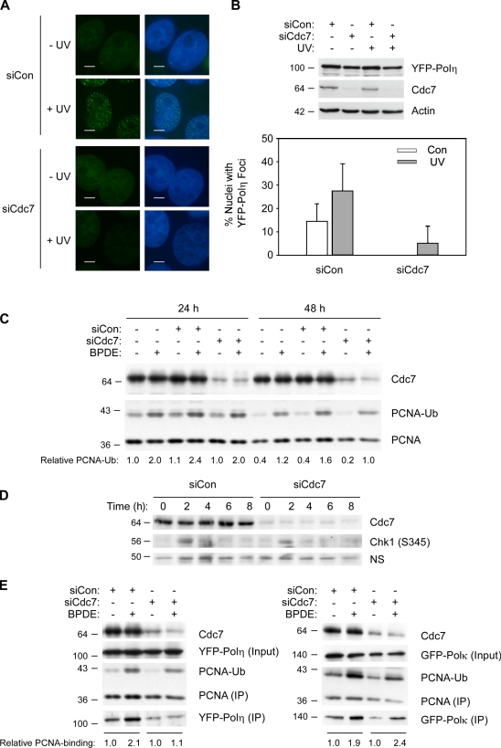
Partial Cdc7 depletion inhibits formation of YFP-Polη nuclear foci. (A) Effect of partial Cdc7 depletion on subcellular distribution of YFP-Polη. YFP-Polη–expressing H1299 cells were transfected with siCdc7 (or siCon) oligos. Some cultures were treated with 100 J/m2 UV. 6 h after UV treatment, cells were fixed and stained with DAPI. Nuclear YFP-Polη foci were visualized by fluorescence microscopy as described in Materials and methods. Bars, 5 µm. (B) Validation of Polη expression and Cdc7 depletion (top). Cell extracts from the experiment described in A and B were analyzed by SDS-PAGE and immunoblotting with anti-GFP (to detect YFP-Polη), anti-Cdc7, and β-actin. Nuclear foci observed in the experiment described in A were enumerated as described in Materials and methods (bottom). To calculate the percentage of cells with Polη foci, ∼250 nuclei were scored for each condition. Data points represent the mean for 250 cells, with error bars representing the range. (C) Effect of Cdc7 depletion on PCNA mono-ubiquitination. H1299 cells were transfected with siCon or siCdc7, or were left untransfected. After 24 and 48 h, the resulting cells were treated with 500 nM BPDE for 1 h. After harvest, cell extracts were analyzed by SDS-PAGE and immunoblotting with the indicated antibodies. (D) Effect of Cdc7 depletion on kinetics of Chk1 phosphorylation. H1299 cells were transfected with siCon or siCdc7. After 24 h and 48 h, the resulting cells were treated with 500 nM BPDE and harvested at the indicated times. Cell extracts were analyzed by SDS-PAGE and immunoblotting with the indicated antibodies. (E) Effect of Cdc7 depletion on PCNA binding of Polη and Polκ. YFP-Polη– or GFP-Polκ–expressing H1299 cells were transfected with siCon or siCdc7. 24 h later, cells were treated with BPDE for 4 h or left untreated for controls. Chromatin fractions from the resulting cells were immunoprecipitated with PCNA antibodies, and the resulting immune complexes were analyzed by SDS-PAGE and immunoblotting with the indicated antibodies. Relative levels of YFP-Polη and GFP-Polκ in the PCNA immune complexes were quantified by densitometry and are expressed as fold change relative to controls (that received no BPDE). Molecular mass is indicated in kilodaltons next to the gel blots.

