Figure 2.
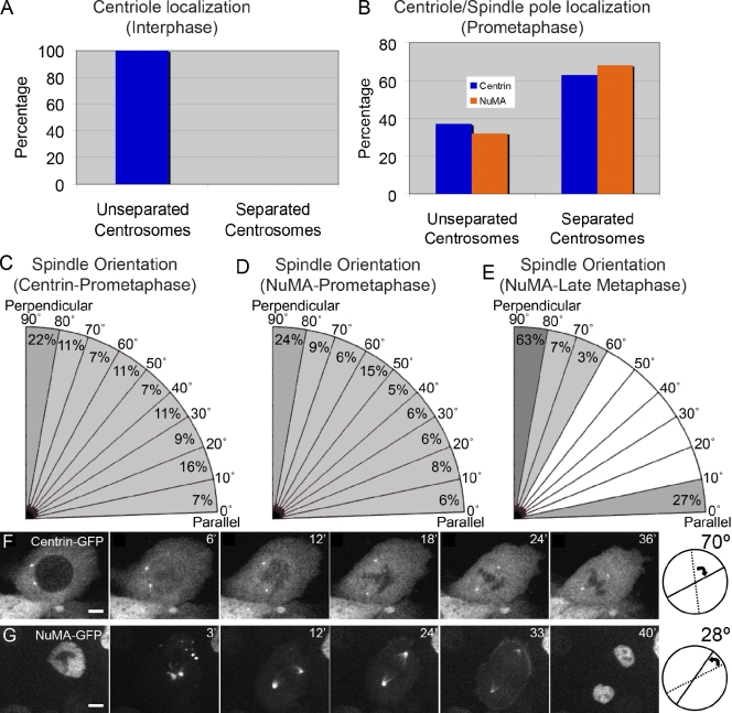
Cell division orientation is established during metaphase. (A) Quantification of basal epidermal cells in interphase with unseparated and separated centrosomes as marked by centrin (n = 1,012 cells). (B) Quantification of basal epidermal cells in prometaphase with unseparated and separated centrosomes/spindle poles. Centrosomes were identified by centrin-GFP localization, and spindle poles were identified by NuMA-GFP localization (n = 98 cells for centrin-GFP; n = 132 for NuMA-GFP). (C–E) Spindle orientation was determined in cells with bipolar spindles as visualized with centrin-GFP in prometaphase (C), with NuMA-GFP in prometaphase (D), and with NuMA-GFP at late metaphase (E). Angles are relative to the basement membrane. (F and G) Rotation of mitotic spindles in cultured keratinocytes as visualized with centrin-GFP (F) and NuMA-GFP (G). (right) Diagrams indicate the angle of rotation of the spindle. Bars, 2.5 µm.

