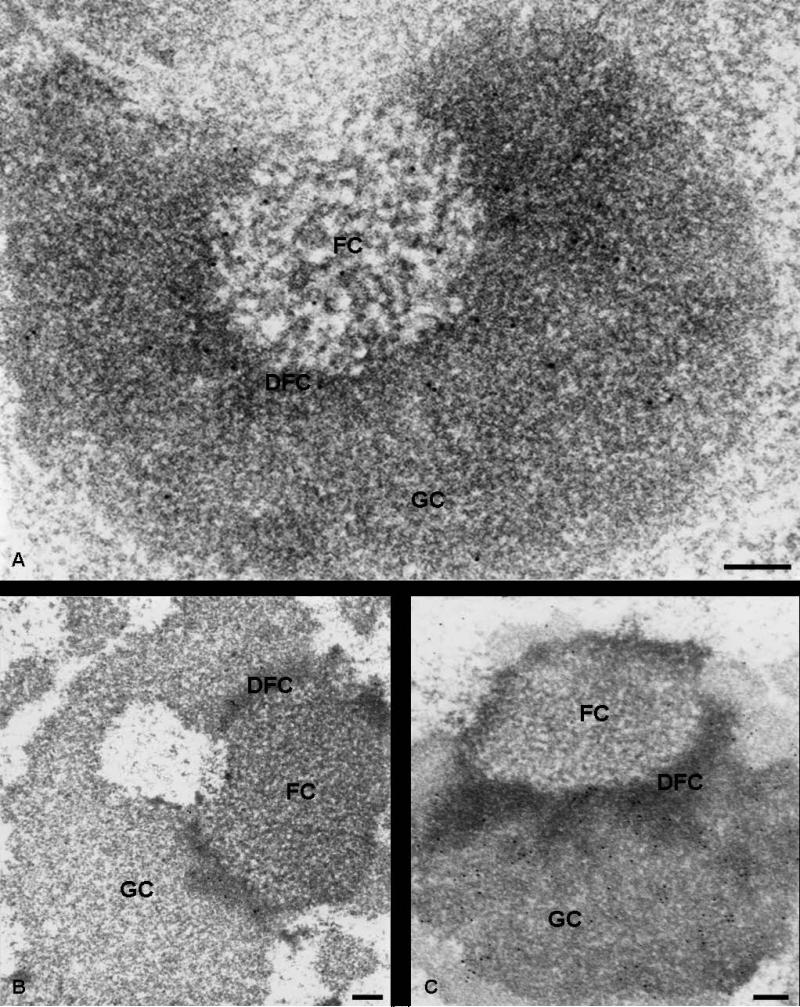
Fig. 3.
Immunoelectron microscopy localization of Nopp140 (a), fibrillarin (b) and nucleolin (c) within the nucleolus in cells treated with actinomycin D. Ultrathin sections of ELT cells, treated for 2 h with 0.05 μg/ml actinomycin D, were immunolabelled with anti-Nopp140 (RE10, a), anti-fibrillarin (b) and anti-nucleolin (c) antibodies. (a): in the segregated nucleolus, labelling was clearly evidenced over the FCs and the DFC, while the GC displayed only a few rare gold particles. (b): the DFC of the segregated nucleolus is the only compartment labelled by anti-fibrillarin antibodies. No labelling was observed over the FCs and the GC. (c): both the DFC and the GC were labelled by anti-nucleolin antibodies, while the FC was devoid of particles. Bars are 0.2 μm

