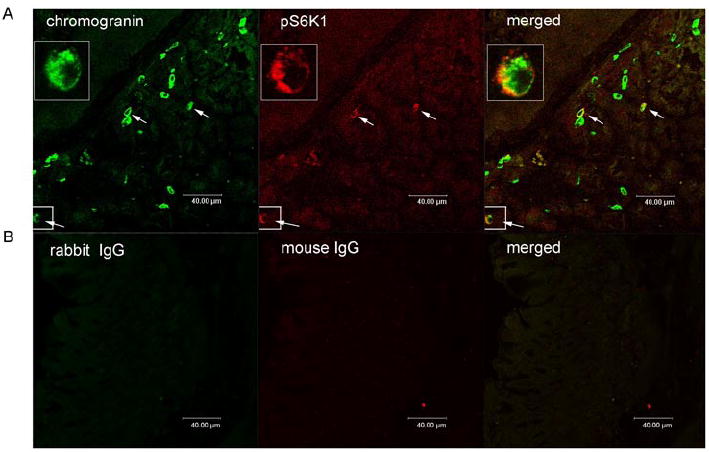
Figure 2. Co-localization of pS6K1 with chromogranin A.

Images depicting chromogranin A (green) and phosphoS6K1 (pS6K1) (red) in gastric mucosal cells. Merged image illustrates co-localization of phosphoS6K1 and chromogranin A (orange) (panel A). Controls included substituting primary antibodies with mouse IgG and rabbit IgG (panel B). Cells expressing both pS6K1 and chromogranin A were marked with white arrows.
