Fig. 4.

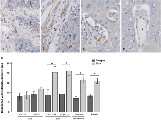
Blood vessel density. Pictures show representative immunohistochemical CD31 staining of a skin tumor and b tongue tumor of the oral SCC cell line HSC-4. CD31 staining is shown in brown and cell nuclei are stained blue. Tumor and stromal areas are indicated with t and s, respectively. Original magnification, 100×. c CD31-positive blood vessels within intratumoral stromal tissue of a tongue tumor of the HSC-4 cell line. Original magnification, 400×. d CD31-negative blood vessel within the same tumor as in c). Blood vessel without CD31 staining is labeled with asterisk. Original magnification, 400×. e Graph shows mean blood vessel density (MVD; ±SEM) of tongue (black bars) and skin (grey bars) tumors of the different cell lines, calculated by counting lumens containing red blood cells. N = 5 for SCC-25, UT-SCC-12A, UT-SCC-7 and Ishikawa tumors. N = 3 for skin tumors and N = 5 for tongue tumors of the HSC-4 cell line. N = 25 for pooled tongue tumors, and N = 23 for pooled skin tumors of all cell lines. *P < 0.05
