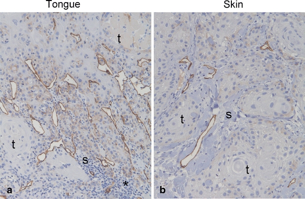
Fig. 8.

Lymphatic vessels in human SCCs. Pictures show representative immunohistochemical staining with antibody against podoplanin (D2-40) of tumor tissue from patients with tongue SCC (a) and skin SCC (b). Podoplanin staining is shown in brown and cell nuclei are stained blue. Tumor and stromal areas are indicated with t and s, respectively, asterisks indicate area with prominent inflammatory infiltrate. Original magnification, 100×
