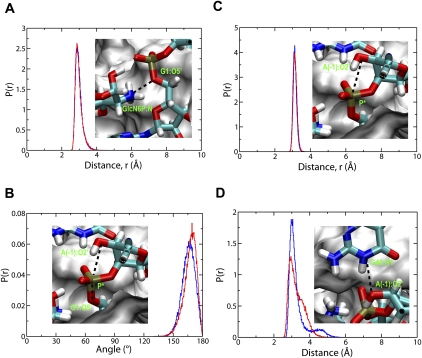
FIGURE 3.
Normalized probability distribution, p(r), of important atomic distances and angle in the binding site of both the wild-type (blue lines) and G40A inactive mutant (red lines) of the glmS ribozyme. (A) Distance from GlcN6P:N to G1:O5′. (B) Distance from A(-1):O2′ to scissile P (P*). (C) Angle between A(-1):O2′, P*, and G1:O5′. (D) Distance from G40:N1 to A(-1):O2′ (A40:N1 to A[-1]:O2′ in the G40A inactive mutant). The insets in A to D are the atomic view of these distances and angles. The cleavage site A(-1) and G1, the metabolite GlcN6P, and residue 40 are shown and colored by element. The rest of the active site is shown as white surface. The functional essential atoms are labeled (green) within the figure.

