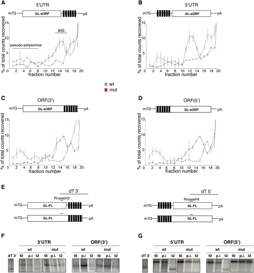
FIGURE 2.
miR-2 controls translation initiation and triggers deadenylation from both UTR- and ORF-binding sites. (A–D) The radiolabeled GL-sORF reporters were incubated in the Drosophila cell-free extracts in the presence of cycloheximide. At the end of the reaction, they were resolved through 15%–45% sucrose gradients, fractionated, and analyzed by scintillation counting. On all wt reporters, inhibition of 80S complex assembly (fractions 10–15) and the formation of pseudopolysomes (fractions 1–8) can be detected. Shown are averages and standard deviations from three independent experiments. (E) Schematic representation of the oligonucleotides used in the deadenylation assays. The dT 3′ and dT 5′ fragments were obtained by annealing the reporter mRNAs to both an RnaseH and a dT oligo and subsequent digestion with RNase H. Therefore, they serve as size markers for the identification of deadenylated reporter mRNAs. Both the wt reporters bearing the miR-2-binding sites at the 3′ end (F) and at the 5′ end (G) are deadenylated at the end of the repression assay (t2), while mut RNAs maintain full poly(A) tail lengths. No change in the adenylation status is observed after the preincubation (p.i.) step. t0 represents input samples at the beginning of the repression assay. The data shown are representative of three independent experiments.

