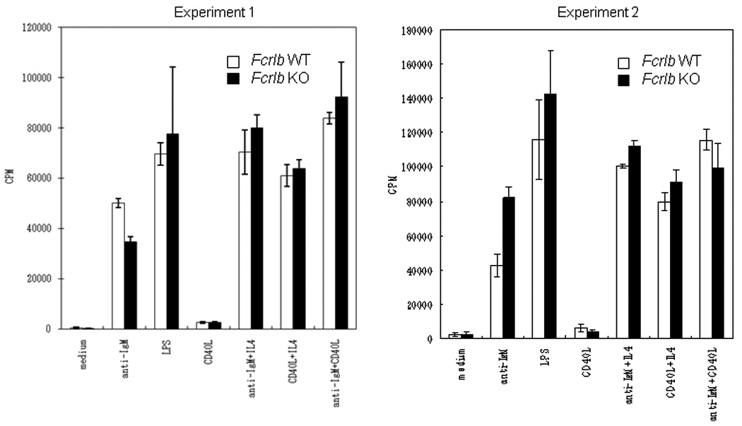
Fig. 3. In vitro B cell proliferation.

Purified splenic B cells were stimulated with anti-IgM, LPS, and CD40L, as well as with the indicated combinations of these stimuli. The proliferative response of KO and WT B cells was analyzed at 48 hours. Data from two independent experiments, each done with a pair of WT and KO mice are depicted. The bars indicate the standard deviation of four assay wells.
