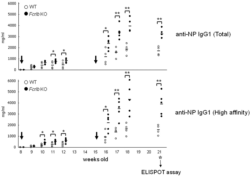
Fig. 4. Primary and secondary antibody responses after immunization with NP16CGG.

Mice were immunized when they were 8 weeks old and boosted at 15 weeks (indicated by the large arrows) with alum precipitated NP16CGG. Serum samples were collected at the indicated time points for analysis of total and high affinity NP-antibodies by an ELISA assay. Six weeks after the second immunization the mice were sacrificed and antibody secreting cells in spleen and bone marrow were enumerated by an ELISPOT assay (Fig. 5). Each symbol represents one mouse. *, p < 0.05; **, p <0.01 (unpaired t-test).
