Figure 1.
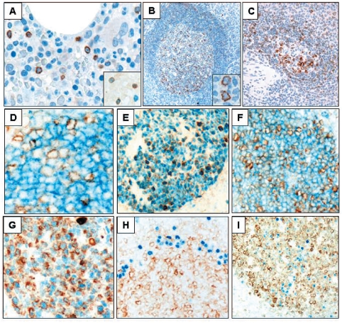
The expression of VpreB3 (brown) in human hematopoietic and lymphoid tissues. Bone marrow biopsies (A) showing, scattered VpreB3 positive lymphoid cells (hematoxylin counterstain for nuclei, 400x), and (inset) partial co-localization of VpreB3 with the lymphoid immaturity marker TdT (dark blue nuclear, no hematoxylin counterstain, 1000x). Reactive tonsil (B) showing VpreB3 staining in a subset of lymphoid cells concentrated within germinal centers (hematoxylin counterstain, 100x, inset 1000x). Spleen (C) showing VpreB3 staining in a subset of lymphoid cells within the white pulp (hematoxylin counterstain, 100x). Tonsil (D-I) co-stained for VpreB3 (brown) and (D) CD10, (E) BCL6, (F) HGAL, (G) LMO2, (H) MUM1/IRF4, and (I) BLIMP1 (blue stains, no hematoxylin counter-stain, D and G at 1000x; E, F, H and I at 200x).

