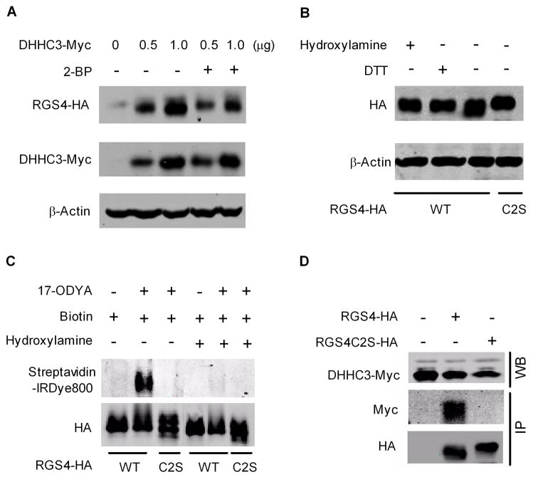
Figure 3.
DHHC3 palmitoylates RGS4 in HEK293 cells. Cell lysates were analyzed by western blot. Images are representatives of 3–6 separate experiments. (A). DHHC3 increased RGS4 protein in a dose-dependent manner, which was blocked by 2-BP (100 μM). (B). DTT or hydroxylamine reversed DHHC3-induced increase in the mobility of RGS4. (C). HEK293 cells co-transfected with GFP-tagged DHHC3 and RGS4 or RGS4C2S were metabolically labeled with 17-ODYA. RGS4 and RGS4C2S were immunoprecipitated and biotin labeled. Samples were treated with Tris-HCl (lanes 1–3) or hydroxylamine (lanes 4–6) for 1 hr prior to western blot analysis using anti-HA antibody and streptavidin-IRDye800. (D). DHHC3 forms complex with RGS4 but not RGS4C2S in HEK293 cells. Expression of DHHC3-Myc was analyzed by western blot (upper panel). HA-tagged RGS4, RGS4C2S, and associated DHHC3-Myc were immunoprecipitated using anti-HA affinity agarose beads and detected by western blot using anti-HA and anti-Myc antibodies, respectively (lower panel).

