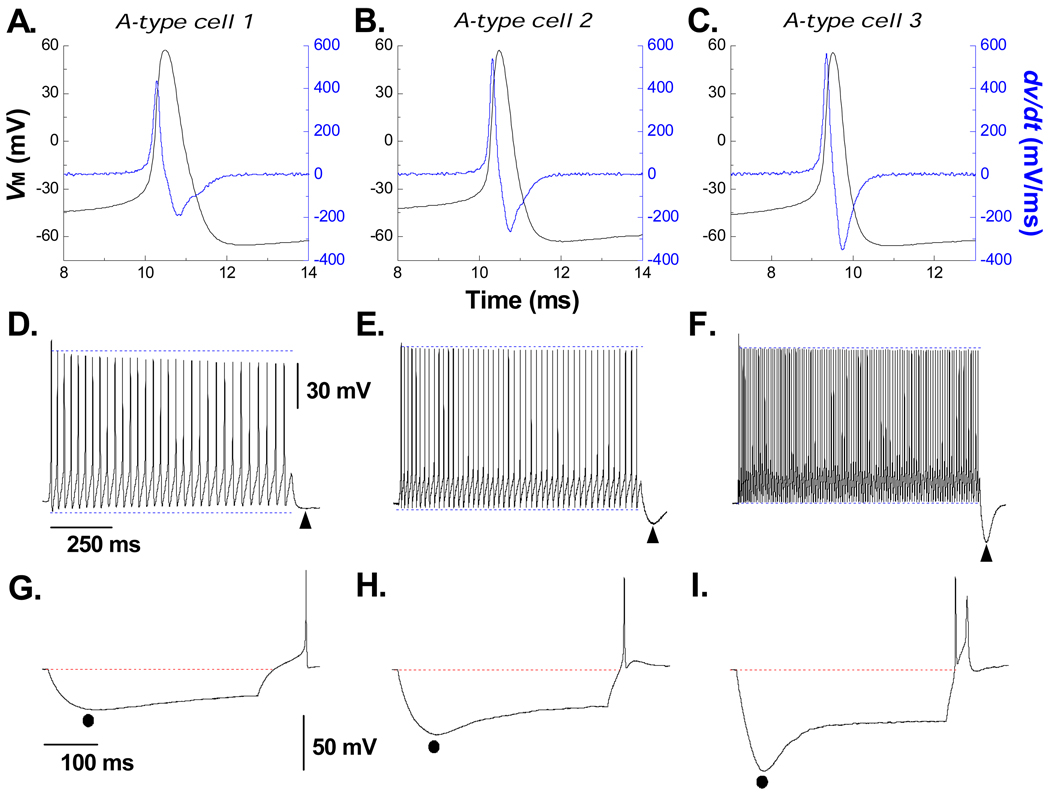
Figure 1. Myelinated A-type vagal neurons express varying levels of cell excitability and hyperpolarization-induced depolarizing voltage sag.
Three distinct examples from a population (n = 47) of myelinated A-type vagal neuron (VGN) implicating that increased cell excitability generally presents with an increased magnitude and time course of hyperpolarization-induced depolarization voltage sag (DVS). A) Single action potential (AP, black trace) evoked by a brief current pulse along with the corresponding dv/dt (blue trace). B) Sustained repetitive AP discharge in response to a 1000 msec, 150 pA current step. Immediately upon termination of the step a period of post excitatory membrane hyperpolarization was observed (PEMH, ▲). C) In response to a 400 msec, −120 pA current pulse membrane potential reached a peak hyperpolarization (●) followed by a depolarizing voltage sag (DVS). Immediately upon termination of the hyperpolarizing step current the trajectory of membrane potential back toward the RMP often elicited an AP. Protocols were the same for the two other example VGN (D – F and G – H) but with a clear trend toward an increased excitability revealing a more prominent magnitude and time course of the PEMH, peak hyperpolarization and DVS.

