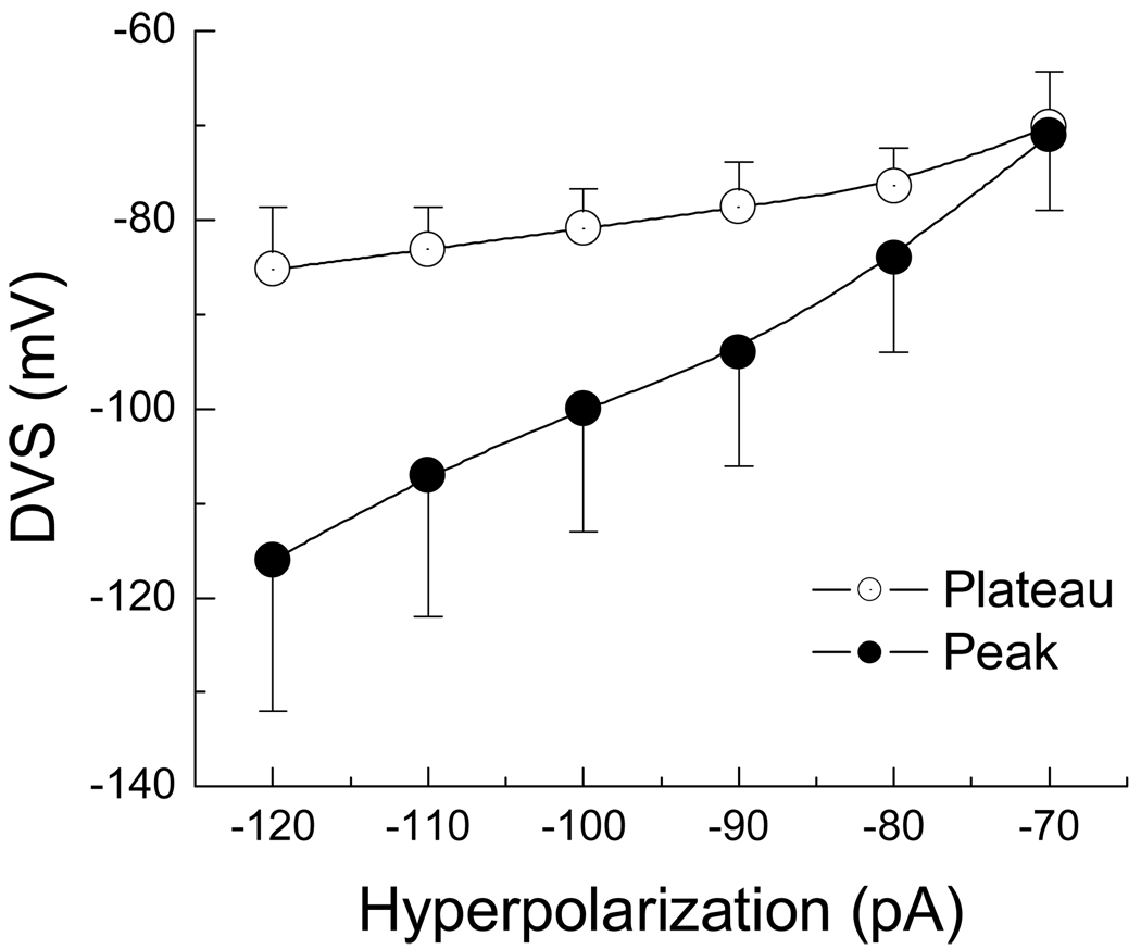
Figure 4. Current-voltage profile of the DVS in A-type VGN.
In a subset of myelinated A-type VGN (n = 6) the peak (●) and plateau (○) membrane potentials were measured in response to 400 msec hyperpolarizing current steps from RMP and presented as an average current-voltage plot (IV). Despite substantial membrane hyperpolarization near the start of the current pulse, the DVS plateaued to membrane potentials within a range of ~10 mV.

