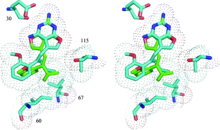
Figure 4.
Superposition of Z3 (isopropyl; green) and E2 (propyl; cyan) in the active site of hDHFR highlighting the different modes of binding for the E and Z isomers of the furopyrimidine analogs. Also highlighted with van der Waals surfaces are the residues that make contact with the inhibitors: Ile60, Leu67 and Val115. Note that the C9-substituents occupy the same conformational space. This figure was generated with PyMOL (DeLano, 2002 ▶).

