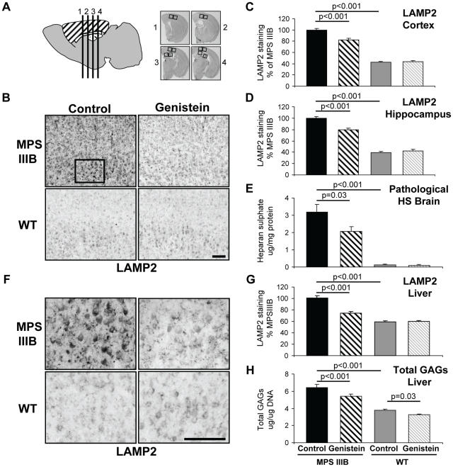
Figure 1. Primary storage substrates are reduced in brains of MPSIIIB mice after genistein treatment.
(A) 11 month old MPSIIIB and WT, male and female mice with and without long-term genistein treatment were sacrificed and 30 µm coronal sections (numbered 1–4) were cut from each mouse at positions 0.26, −0.46, −1.18 and −1.94 relative to bregma. For cerebral cortex (hatched), two low power fields of view for each section (boxed) were quantified or positive cells counted (8 fields total), whilst for hippocampus (spots), two low power fields from sections 3–4 (boxed) were quantified (4 fields total). All sections were stained together and blinded to ensure consistency. (B) Representative Lysosomal Associated Membrane Protein (LAMP2) staining of cerebral cortex of 11 month old MPSIIIB and WT, male and female mice with and without long-term genistein treatment. This indicates the size of the lysosomal compartment and hence stored material in cells in layers II/III-V/VI of the cerebral cortex. The images correspond to section 2 shown in Figure 1A. Bar = 100 µm. (C) Quantification of mean LAMP2 staining in cerebral cortex is expressed as a percentage of staining in untreated MPSIIIB mice. (D) Quantification of mean LAMP2 staining of hippocampus. (E) Mean weight of pathological heparan sulphate in the brain per mg protein, measured using the SensiPro assay. (F) High power view of cerebral cortex layer V - box from (B). Bar = 100 µm. (G) Quantification of mean LAMP2 staining of 2 fields of view from 3 liver sections (6 fields total) is expressed as a percentage of staining in untreated MPSIIIB mice. (H) Mean weight of total glycosaminoglycans in the liver per µg DNA, measured using the Blyscan assay. For all graphs genders were pooled, thus n = 12 mice per group, error bars represent SEM, p values are for Tukey's multiple comparisons test.

