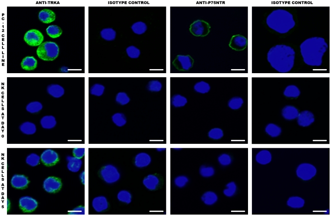
Figure 3. TrkA expression on NK cells as assessed by confocal microscopy.
Cells were prepared for confocal microscopy as described in Materials and Methods. TrkA, but not p75NTR, is expressed by activated NK cells (green staining). On fresh NK cells (NK Day 0), the expression level of TrkA is probably too low to be evidenced by confocal microscopy, in contrast to flow cytometry. PC12 is a rat pheochromocytoma cell line that serves as positive control, as it expresses both TrkA and p75NTR at high levels. The bar corresponds to 3 µm. Data shown are from one representative experiment out of three performed.

