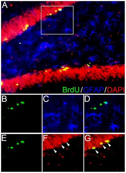
Figure 2. Gliogenesis is minimal in adult hippocampus.
Hippocampal sections were immunostained for BrdU (green), GFAP (blue), and DAPI (red; color changed to red for purposes of visibility) to assess specificity of BrdU labeling and gliogenesis. (A) Representative photomicrograph (400×) of the dentate gyrus of the hippocampus expressing all three labels. Tissue was double-labeled immunofluorescently with antibodies against BrdU (B) and GFAP (C) to determine whether BrdU-labeled cells were glia (D). Approximately 2% of all BrdU-positive cells co-labeled with GFAP. In the triple-labeled image (D), three cells are labeled for BrdU, but do not co-express GFAP. Tissue was also labeled with fluorescent antibodies against BrdU (E) and DAPI (F) to ensure the specificity of BrdU labeling in mature neurons (G).

