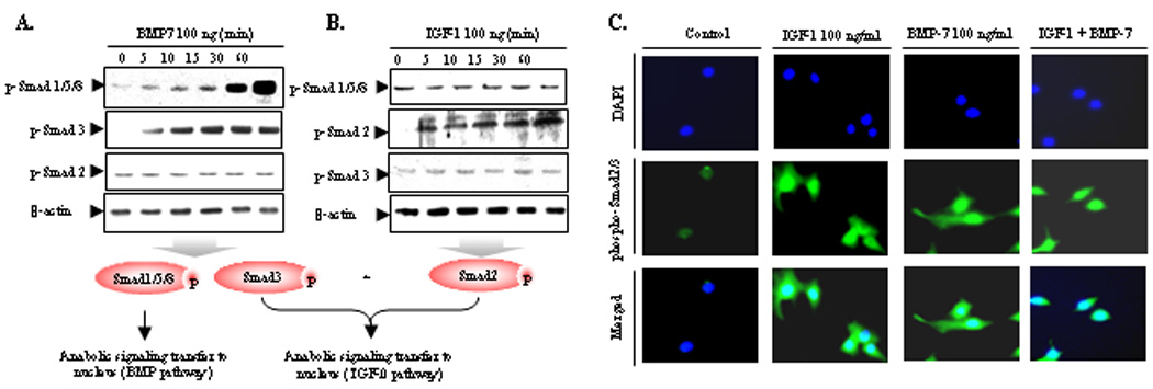
Figure 5.

Combination of IGF-1 and BMP7 activates not only SMAD1/5/8 but also SMAD2/3 mimicking TGF effect. A and B, Serum-starved NP cells were stimulated by IGF-1 (100 ng/ml) and BMP7 (100 ng/ml) for the indicated periods of time. Cell lysates were then prepared and analyzed by western blotting with specific anti-phospho-Smad 1/5/8, anti-phospho-Smad2, and anti-phospho-Smad3 antibody. C, Serum-starved NP cells were cultured in monolayer in 4-well chamber slide for 24 hrs before treatment. Cells were treated with IGF-1 (100 ng/ml), BMP7 (100 ng/ml), and cocktail of IGF-1 and BMP7. 3 hrs later, the cells were fixed with 1% paraformaldehyde, permeabilized in 0.2% Triton X-100. Nonspecific signals were blocked with normal goat serum. NP cells were incubated overnight at 4°C with rabbit anti-phospho-R-Smad polyclonal antibodies followed by incubation with Oregon green®488–conjugated goat anti-rabbit IgG. As a nuclear counterstain, all cells were incubated with 4,6-diamidino-2-phenylindole. The nuclear image was visualized with an ultraviolet filter; the cellular fluorescent stain was visualized with a green filter using a Nikon Eclipse E600 microscope connected to a personal computer, with the results analyzed on MetaMorph Imaging software (series 6.1; Universal Imaging, West Chester, PA). Both images were overlaid with Universal Imaging MetaView imaging software. Three independent experiments in triplicates were performed.
